Patient Stories
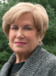
 Patient: Deanne Sherman
December 2011
Deanne Sherman, age 64, West Chester, Pennsylvania
The former celebrity reporter with three sons and four grandchildren was told she may have to have her esophagus removed to prevent esophageal cancer. Then she found out about Cellvizio, the world’s smallest microscope…
Several years ago, when Ms. Sherman was diagnosed with Barrett’s esophagus, a condition of abnormal, potentially precancerous change in the lower esophagus, the initial recommendation from her physician was to have her entire esophagus removed because he couldn’t see the pre-cancerous tissue with the imaging tools he used “I have to find another doctor,” she thought. Ms. Sherman decided to get a second opinion at Lankenau Medical Center in Wynnewood, Pennsylvania where Dr. Bob Etemad told her that she would not have to undergo such a drastic surgery because of a new imaging technology called Cellvizio. With the Cellvizio, Dr. Etemad was able differentiate all the suspicious looking tissue in her esophagus and remove it on the spot with minimally invasive techniques.
“I can’t tell you how relieved I was to hear Dr. Etemad tell me there was another option,” said Ms. Sherman. “Since I retired, my husband and I had made many plans to travel and spend time visiting family and friends. After three minimally invasive endoscopy procedures, I am cancer free and ready to live my life.”
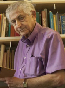
 Patient: Yoram Barzel
November 2011
Dr. Yoram Barzel, a native Israeli and professor of microeconomics at the University of Washington feels he’s a lucky man. Two and a half years ago after a surgery left him suffering from chronic acid reflux, the 80 year-old grandfather of four was diagnosed with Barrett’s Esophagus, a condition in which the lining of the esophagus is damaged by stomach acid, and can lead to cancer of the esophagus.
As recently as seven to ten years ago, the only treatment for Professor Barzel’s condition would have been to remove his esophagus, because imaging technologies weren’t powerful enough to differentiate the potentially cancerous cells from healthy ones. But Dr. Michael Saunders at the University of Washington Medical Center told him that he had another option. During an endoscopy procedure, Dr. Saunders used Cellvizio, the world’s smallest microscope, to see the individual cells lining his esophagus. He was able to target the pre-cancerous tissue and performed a minimally invasive procedure to remove it on the spot. Prof. Barzel returned to Dr. Saunders twice for similar endoscopy procedures and has been deemed Barrett’s and cancer free.
 Patient: Gary Rolf
December 2011
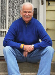
Gary Rolf, age 65, New London, Connecticut
A retried grandfather and life-long nuclear construction worker, Gary battled with esophageal issues related to acid reflux for many years…
Gary had always been very proactive about his health because the amounts of radiation he was exposed to during his career. So, after his primary care doctor told him that his acid reflux could be causing dangerous changes to the lining of his esophagus, he went to go see specialist, Dr. Uzma Siddiqui at Yale Medical Center.
Dr. Siddiqui told Gary that he was in the early stages of Barrett’s Esophagus, a condition that can lead to esophageal cancer.
She and her colleagues were using a new advanced imaging tool called Cellvizio that allowed her to see individual cells during endoscopy procedures.
Instead of taking random biopsies of Gary’s esophagus as physicians have had to do in the past, she used Cellvizio to pinpoint all of the potentially dangerous cells.
After several minimally invasive procedures, he no longer has any remaining Barrett’s tissue. “I can’t stress enough how important it is to monitor one’s health closely. Because of new research tools like the Cellvizio, I can enjoy spending time with my ten grandchildren, attending New York Giants and Knicks games and travelling to the U.K.,” said Gary.
